中国医生实施了一项史无前例的手术,挽救了一名患有快速生长肿瘤的胎儿。
Chinese doctors have performed an unprecedented surgery to save an unborn child who developed a fast-growing tumour.
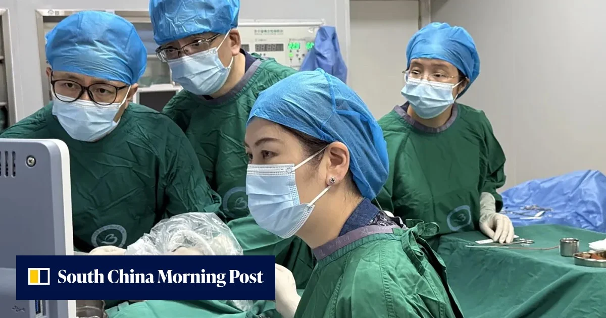
来自深圳和香港的医疗团队最近完成了一项高难度、高精度的胎儿胸腔肿瘤切除术,手术在婴儿仍在子宫内时进行,为面临类似严峻预后的家庭带来了新的希望。
A medical team from Shenzhen and Hong Kong recently completed a challenging, precise removal of a foetal chest tumour while the baby was still in the womb, offering new hope to families facing similarly dire prognoses.
该手术由深圳市宝安区妇幼保健院和香港中文大学的医生联合实施,其详细过程已于8月20日发表在同行评审期刊《产科与妇科超声》上。
Jointly performed by doctors from Shenzhen Baoan Women’s and Children’s Hospital and the Chinese University of Hong Kong, the procedure was detailed and published on August 20 in the peer-reviewed journal Ultrasound in Obstetrics & Gynecology.
据大陆科学媒体平台“先进生命科学”9月15日的一篇文章称,这标志着中国在胎儿实体瘤微创介入治疗领域“迈向了全球前沿”。
This marks China “stepping to the global forefront” in the field of minimally invasive interventional treatments for solid foetal tumours, according to the mainland science media platform Advanced Life Science in a September 15 article.
医疗报告称,这位40岁的母亲在怀孕22周时,其胎儿被诊断为微囊型先天性肺气道畸形(CPAM)。
The 40-year-old mother was 22 weeks pregnant when her foetus was diagnosed with microcystic congenital pulmonary airway malformation (CPAM), the medical report said.
CPAM是一种罕见的先天性疾病,由胚胎发育期间肺组织异常生长引起。虽然它是良性的,但属于快速生长的肺部肿瘤,可能导致心力衰竭和水肿,即严重的液体积聚。
CPAM is a rare congenital condition caused by the abnormal growth of lung tissue during embryonic development. While benign, it is a rapidly growing lung tumour that can lead to heart failure and hydrops, a severe accumulation of fluid.
扫描显示,有一个约5厘米(2英寸)大小的实体肿瘤,占据了胎儿整个右侧胸腔。它严重压迫了心脏和肺部,并已经导致腹水,即腹腔内危险的液体积聚。
Scans revealed a solid tumour measuring about 5cm (2 inches) and occupying the foetus’ entire right chest cavity. It severely compressed the heart and lungs and had already caused ascites, a dangerous build-up of fluid in the abdomen.
标准治疗通常涉及引流大量液体积聚或切断肿瘤的主要血液供应。然而,由于该特定肿瘤由微小囊肿组成且缺乏中央血管,传统方法无效。为了挽救婴儿,医疗团队采用了一种新方法。在高清实时超声成像的引导下,团队精确规划了一条路径。
Standard treatments usually involve draining large fluid pockets or cutting off a tumour’s main blood supply. However, because this specific growth consisted of tiny cysts and lacked a central blood vessel, traditional methods were ineffective. To save the infant, the medical team adopted a novel approach. Guided by high-definition, real-time ultrasound imaging, the team precisely mapped a route.
他们顺利将一根精细的消融针——一种纤细的专用医疗探针——穿过母亲的腹部、子宫壁和胎儿的胸壁,直接抵达肿瘤的核心部位。
They smoothly inserted a fine ablation needle – a thin, specialised medical probe – through the mother’s abdomen, the uterine wall and the foetal chest wall, reaching directly into the core of the tumour.
微波技术通过振动水分子产生高温,从而从内部彻底破坏肿瘤组织。主动加热过程异常迅速,总共仅用了两分钟。
The microwave technology works by vibrating water molecules to create intense heat, essentially destroying the tumour tissue from the inside out. The active heating process was exceptionally fast, taking a mere two minutes in total.
医生们通过监测设备密切观察热量的扩散情况,以确保婴儿的健康器官不受损伤。
Doctors closely watched the heat spread on their monitors to ensure the baby’s healthy organs remained unharmed.
效果立竿见影。手术仅三天后,肿瘤体积显著缩小,积聚的液体也完全消失。
The results were immediate. Just three days after the procedure, the tumour’s volume had decreased significantly and the fluid build-up was completely gone.
婴儿在怀孕37周时通过剖腹产出生,体重为2.9公斤(6.4磅)。出生时,该婴儿在阿普加评分(用于快速评估新生儿身体状况的测试)中获得了满分10分,表明其整体健康状况极佳。四个月大时,该婴儿仍无任何症状,且无需接受任何手术干预。
The infant was delivered via caesarean section at 37 weeks, weighing 2.9kg (6.4lbs). At birth, the baby achieved a perfect score of 10 on the Apgar scale – a quick test used to check a newborn’s physical condition – indicating excellent overall health. At four months of age, the infant remains symptom-free and has required no surgical intervention.
根据《Advanced Life Science》期刊的文章,此次手术的成功不仅是一项极具挑战性的临床救治,也在国际母胎医学史上“树立了一个新的里程碑”。
According to the Advanced Life Science article, the success of the surgery was not only a highly challenging clinical rescue but also set “a new milestone” in the international history of maternal-foetal medicine.